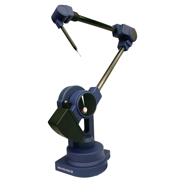
Microscribe CMM used for muscle fiber digitization.

Forearm muscle fiber bundles and tendons with underlying bone surfaces.
[Credit: Zhi (James) Li, Anne Agur, Division of Anatomy, Department of Surgery, Faculty of Medicine, University of Toronto.]
This digitization process is slow and requires patience and dedication. And, before the Parametric Human Project got involved, the process was, unfortunately, even slower. A critical part of the problem was the software. The software that was being used for digitizing the muscle fiber bundles was a hand-me-down from unrelated research projects and had been modified to help out in Anne's lab. Funded by Autodesk Research, the Parametric Human Project started a rewrite of the software tailored specifically for muscle architecture digitization. The new program was called Fibonacci and was designed to match the workflow in the lab. This greatly accelerated Dr. Agur's process: “We had digitized several individual muscles in the past, but when we got Fibonacci, it was dramatically better to the point where we could imagine digitizing the entire human body at a surgical level of detail.“

FIBONACCI Software [Credit: Jacobo Bibliowicz, James McCrae, Azam Khan, Autodesk Research.]
Now, the Musculoskeletal Anatomy Laboratory has digitized over 100 muscles, making significant headway and learning amazing new things about human muscle architecture. Also, the group has taken the digitization of the innervation of the muscles further as well, and have already develop a clinical application for addressing pain in the lower back. Knowing where the nerves are located inside the muscle is also critically important for surgeons when repairing muscle.
As part of the Parametric Human Project network, the muscle digitization work from the University of Toronto has directly helped another lab in their muscle simulation work, across the country at the University of British Columbia. And, although there is still a lot of work to do, completing the muscle architecture map for the entire human body has now become possible, opening up new amazing possibilities.

[Credit: Logo Design by Justin Matejka.]


Coordinate Measuring Machine (CMM)
For a long time, the general understanding of muscle architecture was greatly over simplified. Muscles were considered to have all parallel fiber bundles and when the muscles contract, they become shorter and bulge in the middle. The surprising thing is that there are very few muscles in the human body that work that way. They have complex branches and attachment patterns, have interwoven tendinous sheets, or –like the human tongues– can be a muscular hydrostat, like a tentacle, where the fiber bundles work against each other to produce controlled motion.
Due to this complexity, most muscles in the body have never been mapped out. The arrangement of the muscle fiber bundles, or the muscle architecture, is still unknown for 80% of the 500 human muscles. Luckily, at the University of Toronto, Prof. Anne Agur and her students at the Musculoskeletal Anatomy Laboratory in the Department of Surgery have been making progress on this fundamental problem. By carefully digitizing the 3D shape of each and every fiber bundle along its length, and the related tendinous parts, the group has been creating detailed volumetrically-accurate muscle architecture maps. These beautiful 3D models reveal the amazing complexity of the body and convey some notion of how the muscle would behave in action.