
Distal phalange of the index finger. The top image is the 3D points scanned by the Faro arm laser scanner. The bottom image shows the results of a noise reduction process.
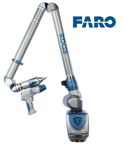

FARO Arm Laser Line Probe (LLP) used to scan bones at the Department of Anatomy, Faculty of Medicine, University of Toronto.
At the University of British Columbia, bone surface scanning was automated with a Coordinate Measuring Machine.


Laser Line Probe (LLP)
Human Skeletal Modeling
To revolutionize parametric skeletal modeling, we employed cutting-edge technology and a multidisciplinary team of experts. The bone scanning project involved the detailed scanning of over 1000 bones, utilizing the Faro arm Laser Line Probe (LLP) 3D Point Cloud scanner to capture point cloud data that was processed with a research system for semi-automated noise reduction and surface meshing. MicroCT was also used on a small sample of finger bones to see if the volumetric data from the bone interiors could be combined with high resolution exterior scans. As the denoising algorithm moves the points toward the statistically likely surface, we also developed a visualization tool to better understand if the point density is consistently high enough for triangulation of the points to create a geometric surface mesh.

Visualization of point cloud density (yellow - high, blue - low).


Micro CT of proximal, middle, and distal phalanges, some of the smallest bones in the human body.
While this technique creates very high quality and high resolution surface models of bones, the interior bone structure is not included. To investigate the data quality we could achieve of bone interiors, we performed micro CT scans of the bones in the fingers. In future, we plan to combine these scans, registering the bone surface meshes to the interior volumetric data.
As the process of laser scanning the bones is quite labour intensive, a Coordinate Measuring Machine (CMM) at the University of British Columbia (UBC) was employed to explore automation for surface scanning. This project resulted the complete scanning of four skeletons, including a plastic reference skeleton to compare results, with multiple scans conducted on a selection of bones to ensure reproducibility.
The use of a CMM for bone surface scanning involves the creation of control macros specifically designed for vertebrae, long bones, ribs, hands, and feet. Each scan, coupled with processing, takes approximately 10-15 minutes, demonstrating the efficiency of the process.

At the Parametric Modeling of Human Anatomy 2014 Conference, hosted by the University of British Columbia, the bone surface scanning Coordinate Measuring Machine process was demonstrated.
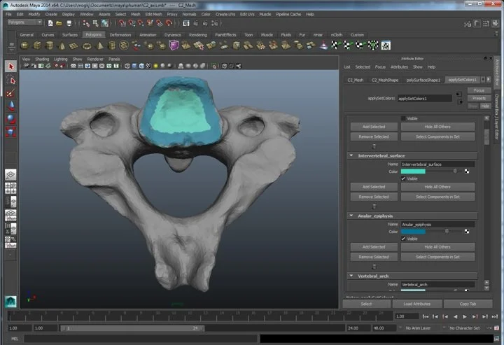
Terminologia Anatomica (TA) is the international standard on human anatomic terminology. It was developed by the Federative Committee on Anatomical Terminology (FCAT) and the International Federation of Associations of Anatomists (IFAA) and was released in 1998. While this taxonomy names many features on bone surfaces, no textual descriptions are given and no imagery is provided. To address this gap, we investigated methods and tools that would be helpful to create a digital version of the TA that we called the Computational Terminologia Anatomica (CTA). In Autodesk Maya, shown above, 3D painting tools were customized so that expert anatomists could paint bone areas that were named in the TA and, to capture bone surface edge features, curvature analysis plug-ins were created.
Putting it all together, we developed a web-based CTA tool to both help with the authoring process, and to serve as an anatomy browsing system.