
Left to Right: Azam Khan, George Church, Andrew Hessel (at the CIFAR-GETx Conference in Toronto, Canada (2018)


The outcome of George Church’s genome (exterior view of cephalic region).
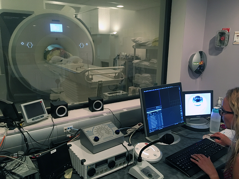


MRI
George Church, Inside Out
Through our colleague Andrew Hessel, we met with Prof. George Church at a number of events, including the GET Conference (founded by George Church). After the Parametric Human Project sponsored the conference in Vienna in 2015 and Boston in 2016, we asked MaRS and CIFAR to help bring the event to Toronto which resulted in CIFAR-GETx Conference 2018.
George Church is a prominent figure in the field of genetics and molecular biology, renowned for his groundbreaking contributions to genomics and synthetic biology. As a professor at Harvard University and a founding core faculty member of the Wyss Institute for Biologically Inspired Engineering, Church has played an instrumental role in advancing our understanding of DNA sequencing, genome editing, and genetic engineering.
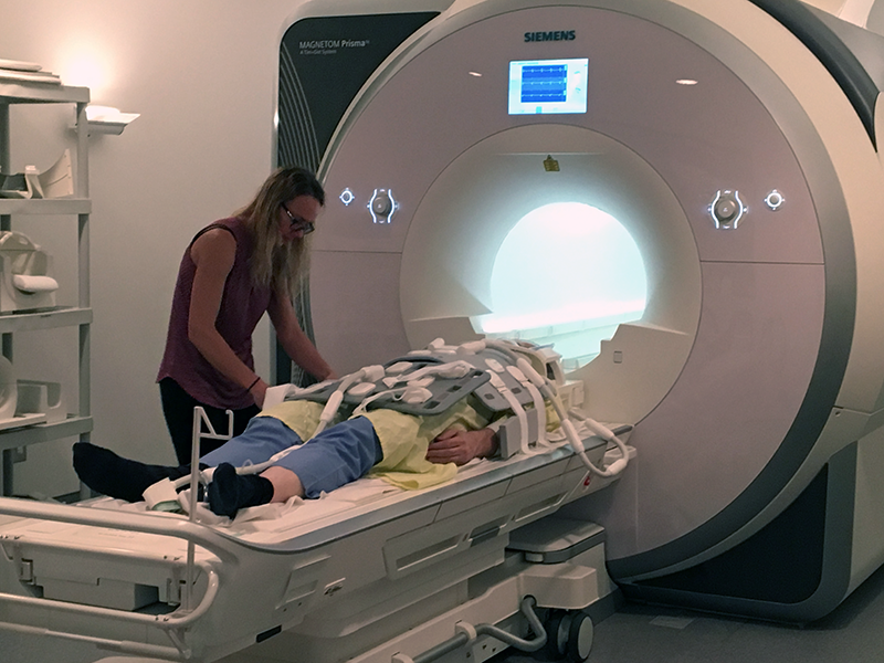
Our particular interest in the initiatives of Prof. Church is his Personal Genome Project (Harvard PGP) which seeks to publicly post complete DNA sequencing datasets of 1,000 people. This effort has led to a global network of national PGP affiliates. This is an incredible contribution to science to have these datasets available at that scale. We think of this as the ultimate set of parameters that can generate any human. However, when we saw how the people associated with each data set were documented, with just a mug shot and a ruler, we saw an opportunity to better match the DNA sequences with a “top down” dataset to better understand what those sequences became. So, the day before the conference in 2018, we took Prof. Church to the research MRI facility at York University in Toronto.
With the help of Joy, at the York MRI Facility, George got strapped down with all the transmit/receive coils. He commented that he felt like the giant in “Gulliver’s Travels” when he was tied down by the Lilliputians. The data from the coils is brought into an Image Reconstruction Computer where radial analog signals are statistically processed into pixels (for 2D images) or voxels (for 3D volumetric datasets).
The purpose of this exercise was to explore what could be done with the system and what quality of imagery could be captured. The ultimate goal would be to combine several scans into one holistic data set that could later be queried for subsets: either Cartesian sub-regions, extracted objects (e.g., bones, muscles, or organs), or types of data, including geometry or time-based imagery.
With a specimen as large as George, we already knew the arms would not fully be captured, nor would the lower legs or feet. For those, we would have to turn George around and send him back into the MRI machine for additional scans, to be aligned and combined later.

The entire MRI system had been upgraded, since we had been there before to scan Andrew Hessel. So, in addition to the imagery we got before, we saw surprisingly detailed brain and heart images.
To the right, from the control room, we start to get the first images of George as he enters the MRI, inside out! You can see a lot of the skeletal muscle, the brain, and how the arms are a bit cropped as they fall outside the range of the coils and imager.
Below on the left, in the midsagittal view, you can see the Corpus callosum, the Limbic lobe, the Hypothalamus, the Medulla oblongata, the Cerebellum, and part of the spine. On the right, we can see some key features of the brain’s vascular system. The brightest spots are the internal carotid arteries forming part of the Circle of Willis.

The heart was especially exciting as an animation could be produced by sampling a specific point in time (a “frame”) during each beat. Each sample would be just a little later in the heartbeat cycle than the previous time. But because the heartbeat cycle is virtually identical for each beat, any frame will look the same as the corresponding frame from the previous cycle.
By having each frame be just a little later than it was before, the animation looks like it is a single heartbeat even though it was combined from a hundred beats.
And the result is truly amazing. You can see the valves opening and closing, the blood flowing in and out of the chambers, and the thickening of the walls of the cardiac muscle.