
Photo rig at Quantum Capture, Toronto. [Image credit: Andrew Hessel]


Photogrammetry close-up of Andrew Hessel.
The photogrammetry software analyzes the photographs to identify common features or points visible in multiple images. These features could include distinctive points on the person's body, clothing details, or any other recognizable elements.
Based on the identified features and camera calibration, the software creates a point cloud – a set of 3D points representing the surface of the person. This point cloud is a raw representation of the object's geometry. The point cloud is then used to generate a dense mesh that represents the surface of the person. The mesh consists of interconnected triangles, forming a detailed 3D model. Finally, the color information from the original photographs is mapped to the surface mesh, creating a textured 3D model that closely resembles the appearance of the person.
This project was highly beneficial in helping to create a protocol for repeating, and improving, future scans. Also, the datasets collected will help us to develop tools to combine these models, ideally, in an automated fashion.
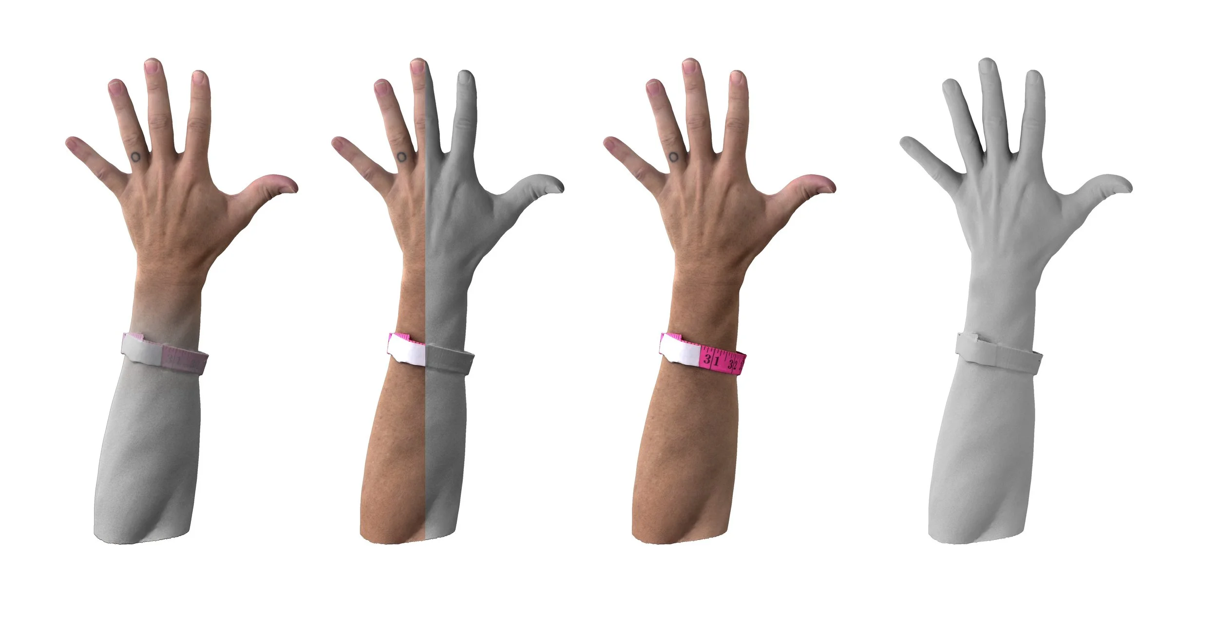
Photogrammetry close-up of Andrew Hessel’s hand.
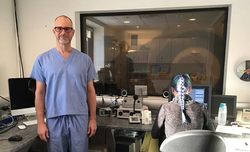
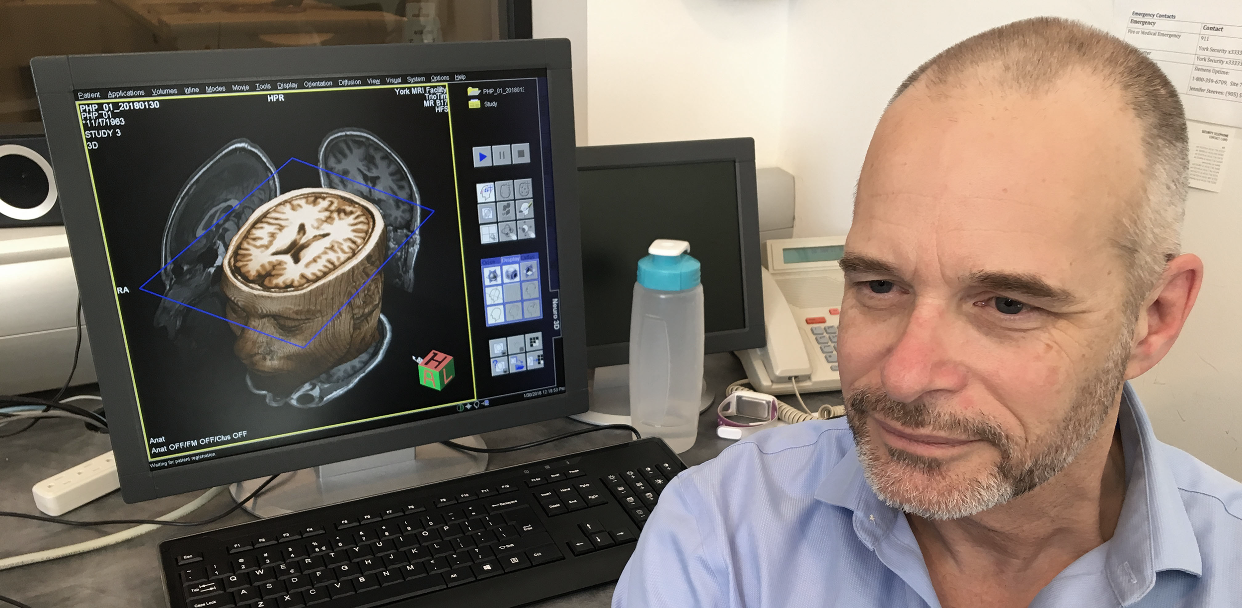
At the York MRI Facility at York University in Toronto, with the help of Prof. Jennifer Steeves and Joy Williams, MRI Technologist, we collected a number of scans of Andrew to create the interior model. In future, we hope to take this combined model and map features to his DNA sequence as well, as Andrew was participant No. 10 in the Personal Genome Project: Canada.
The entire process occurred over the course of a morning session. To collect an internal image of the whole body, several sectional scans are taken. Each scan covers 30 to 40 cm of the subject, with generous overlap between sections, for a registration algorithm to combine these image blocks into a complete whole body MRI. Some preparation is needed to stabilize the subject, to minimize motion while in the scanner. From the control room, instructions and updates are given to the subject.

Unfortunately, when Andrew is laying down in the MRI scanner, he will not be the same shape as he was when the photogrammetry capture was done. To map the surface scan to the volume scan, corresponding features between the two will have to be labelled so that a registration process can deform the surface to the volume. Then, these two data sets together will form the context for more specific scans.
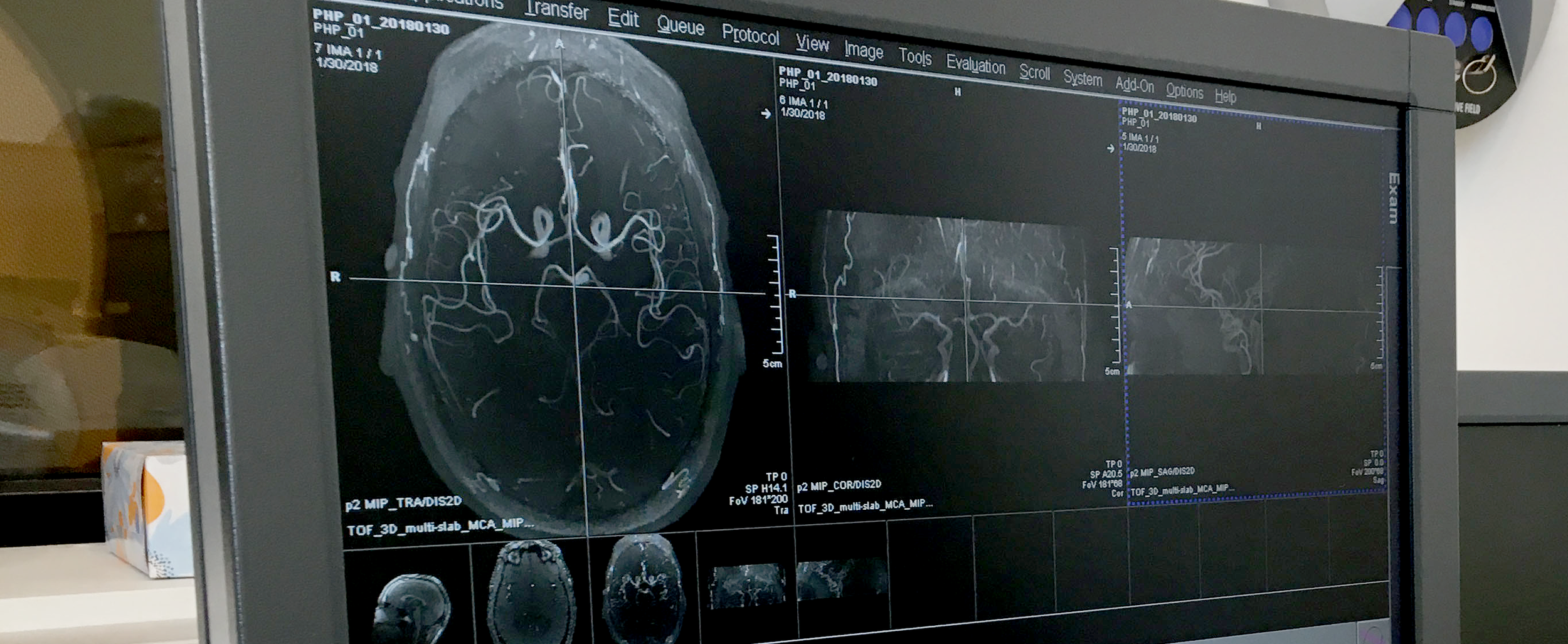
The first scan, a rough overview, is done quickly. And we have our first glimpse inside the body. The result is instantly impressive showing detailed human anatomy. For example, in the sagittal view, the spine comprises about half of the width of the body.
The MRI system has a vast array of parameters and settings. Many settings can be used to reveal different properties of various tissues in the body. For example, Magnetic Resonance Angiography (MRA) is a group of techniques used to clearly image blood vessels, especially in the brain and around the heart.
By combining computational techniques with MRI techniques for estimating the diffusion, or restriction, of water molecules in the tissues and fluids, directions of fibrous tissue can be calculated. This is now common in brain imaging to better understand how different parts of the brain relate to each other. In the scanning software, seed voxels can be selected and the system will trace extracted directions to show fiber traces.
We now have a significant task ahead to determine how, and what tools need to be created, to combine the volumetric MRI data with the geometric surface scan dataset, as well as projecting the muscle fiber bundle dataset into the volumetric data. Another major task is the segmentation of the voxel data into specific bones, organs, and blood vessels. Once these tasks are underway, we can begin the key work in the Parametric Human Project of correlating the data and the extracted features with the relevant sections of the DNA sequence and, later, between Andrew and other subjects.
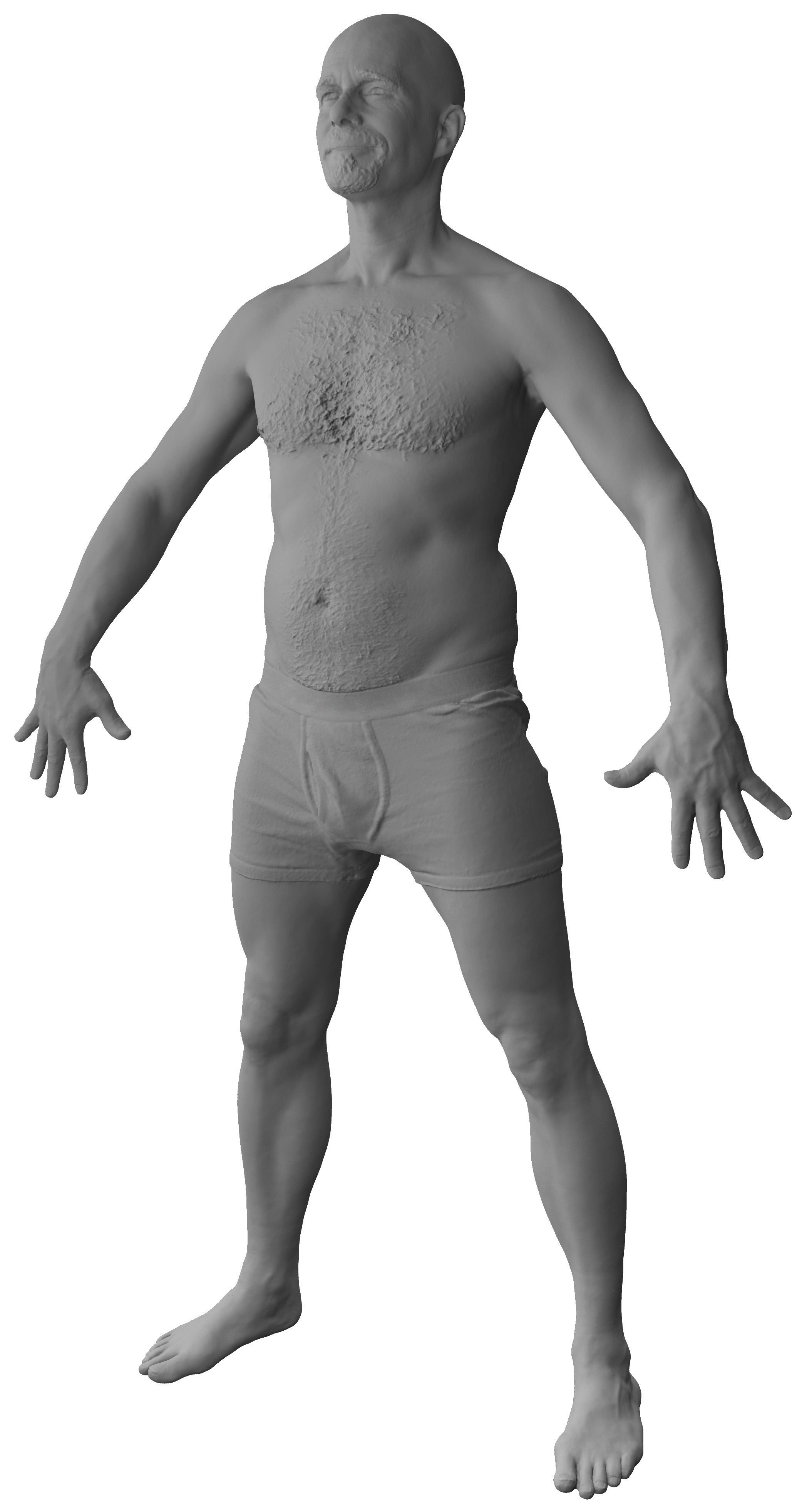
Whole body photogrammetry of Andrew Hessel.


Seeing Andrew from the inside and the outside simultaneously is quite novel, which begs the question, can we bring the data to life to match the living data source?
Photogrammetry
To develop a precise and comprehensive human biomechanical model, it is imperative to capture the intricate details of the interior musculoskeletal system. One method to achieve this is through the use of Magnetic Resonance Imaging (MRI), which is capable of capturing the primary structures within the body. However, despite its efficacy in capturing internal details, the resolution of MRI scans tends to be relatively low. This limitation becomes particularly relevant when aiming for a detailed representation of the external surface of the human body.
To overcome the resolution constraints of MRI scans and achieve a more detailed representation of the external surface, a high-quality surface scan becomes essential. By employing photogrammetry, it becomes possible to create a high-resolution geometric surface mesh of a person. This method allows for a more accurate and detailed depiction of the external features, contributing to the creation of an aggregate biomechanical model that combines the internal and external aspects of the human body with enhanced precision. Through this combined approach, the biomechanical model can better serve various applications, ranging from medical research and diagnostics to animation and virtual simulations.
Photogrammetry is widely used in fields like archaeology, architecture, and entertainment for its ability to create accurate and realistic 3D models from photographs. For human models, it has been used for areas such as virtual clothing try-on, character modeling for games and movies, and even medical applications for creating personalized prosthetics or orthotics. To see how this technique could be used in the Parametric Human Project, we visited Quantum Capture in Toronto with Andrew Hessel.
Photogrammetry is a technique used to create detailed 3D models or surface meshes of objects by analyzing multiple photographs taken from different angles. This involves capturing a series of photographs of the individual from various viewpoints and then using specialized software to reconstruct a three-dimensional representation. Show here is the set-up at Quantum Capture, with 112 DSLR (digital single-lens reflex) cameras. As a test, we tried three set-ups: full body, head, and hand. The purpose of having close-ups of the head and hand was to have higher resolution in these areas, and to later replace those areas of the full body model.